INTRODUCTION
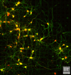
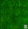
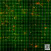
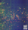
We present 6 different image datasets acquired by fluorescence microscopy and depicting hippocampal in-vitro neuronal networks. The datasets are used for the evaluation of our approach for the joint analysis of neuronal anatomy and functionality, involving the use of a high-resolution Multi-Electrode Array (MEA) technology for the acquisition of the functional signal. Results concerning both the neuronal nuclei detection and the structural/functional representation of the neuron/electrode mapping are provided.


